Questions?
Talk to an Expert
Talk to an Expert
If you're looking for something, we can help! Give us a call at 1 (888) 228-7564 or shoot us an email anytime: Sales@IntegrisEquipment.com

GE MAC 3500 RESTING ECG ANALYSIS SYSTEM Display Type: Backlit 165 mm (6.5 in) diagonal graphics AM LCDPower Source: 100/240 V AC/ Battery: 18 V NiC...
View full details
GE Marquette MAC 5000 EKG Machine Purchase Includes: Refurbished GE (Marquette) MAC 5000 EKG Machine Patient Cables Certified Biomed Equipment Dia...
View full details
GE MAC 5500 ECG Machine Built on GEs proven innovation in ECG acquisition and analysis, the MAC 5500 is GEs premier ECG system, delivering advanced...
View full details
GE MAC 1200 EKG Machine The MAC 1200 EKG Machine from GE Medical is in its basic version a resting ECG system whose true one-button operation mak...
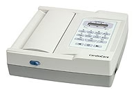
View full detailsBionet CardioCare 2000 Interpretive 12 Channel Resting ECG Part Number: ECG-2000Purchase Includes: CardioCare 2000 Main Unit 10 Lead ECG Patient C...
View full details
Burdick Atria 3000 Interpretive ECG-EKG Machine Simple to use, compact and reliable, the Burdick Atria 3000 ECG is the ideal choice for the physici...
View full details
Burdick Atria 3100 ECG Monitor Part Number(s): A31-1EI01 (Interpretive), A31-1EK01 (Non-Interpretive)Burdick Atria 3100 ECG technology assures h...
View full details
GE Mac 800 Electrocardiograph Optional LAN communication to CardioSoft and MUSE systems The GE MAC 800 EKG enables healthcare providers to connect ...
View full details
Burdick Atria 6100 ECG Monitor Part Number(s): A61-1EI01 (Interpretive), A61-1EK01 (Non-Interpretive), A61-1RI01 (Interpretive with wireless)The...
View full details
QRS Diagnostic PC-Based Universal ECG Electrode Part Number: Z-7000-0301 Turns any PC into a full-featured electrocardiograph. Intuitive Windows op...
View full details
Edan Three Channel Wide Screen ECG Machine Part Number: SE-3BPurchase Includes: Edan SE-3B Three Channel Wide Screen ECG Machine ECG Cable (4mm, b...
View full details
GE MAC 1600 Resting ECG System Optional Internal Memory for storage of 100 ECG'sPurchase Includes: GE MAC 1600 Resting ECG System AHA Multilink Pa...
View full details
GE MAC 1200 EKG Machine 12SL InterpretationBrand New GE MAC 1200 comes with manufacturer's warranty Enhance decision making efficiency and continui...
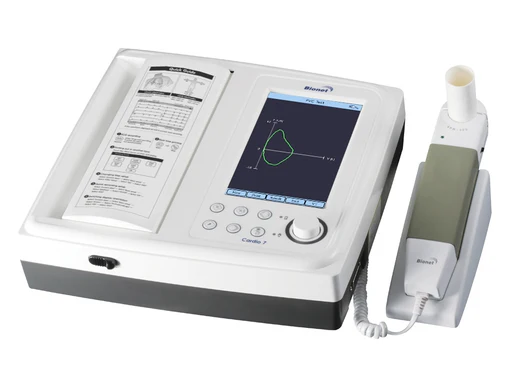
View full detailsCardio7-S 7" Color TFT LCD 12 Channel Interpretive Resting ECG and Spirometer with Touch Screen and WiFi Part Number: Cardio-7BSPurchase Includes: ...
View full detailsCardio7-S 7" Color TFT LCD 12 Channel Interpretive Resting ECG and Spirometer with Touch Screen and WiFi Part Number: Cardio-7BDSPurchase Includes:...
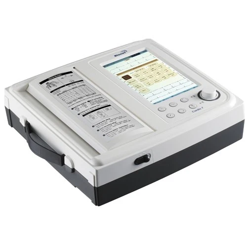
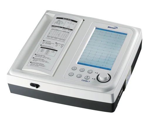
View full detailsCardio7 7" Color TFT LCD 12 Channel Interpretive Resting ECG with Touch Screen and WiFi Part Number: Cardio-7B12 Channel Interpretive Resting ECG w...
View full detailsCardio7 7" Color TFT LCD 12 Channel Interpretive Resting ECG with Touch Screen and WiFi Part Number: Cardio-7BPurchase Includes: Cardio7 Main Unit...
View full details
Philips PageWriter Trim I Cardiograph Part Number: (860290) High performance without high costs For unmatched patient convenience, the Philips Pag...
View full details
Philips PageWriter Trim III Cardiograph Part Number: 860286 Get connected with PageWriter Trim III Does your organization store ECGs in a central ...
View full details
The E-12 ECG Machine by Embra Medical - Competitively Priced EKG Machine for Sale EMBSE-1200EB Embra Medical is committed to providing an advance...
View full details
Z Fold ECG Recording Paper for Burdick Atria, 1 Pack Part Number: 007983 Chart Type: Z-Fold (Fan fold) 200 SheetsDimensions: 8.5 x 11 inchGrid Colo...
View full details
Burdick Atria 6100 and 3100 ECG Power Supply Part Number: 010-1684-00Application: Power supply for use with Burdick Atria 3100 and Burdick Atria 61...
View full details
Burdick 6100 ECG Patient Cable Part Number: 012-0844-01Specifications: 10 Pin Leads, AHA Patient Cable Burdick 10L EKG Resting Cable, Banana Plug N...
View full details
Burdick Cardiosens Ultra II ECG Electrodes - 100/Box Part Number: 047029 Electrodes consistantly provide high quality tracing by ensuring that each...
View full detailsWhat is an EKG, or what is an ECG?
Electrocardiography (ECG or EKG from the German Elektrokardiogramm) is a transthoracic (across the thorax or chest) interpretation of the electrical activity of the heart over a period of time, as detected by electrodes attached to the outer surface of the skin and recorded by a device external to the body. The recording produced by this noninvasive procedure is termed an electrocardiogram (also ECG or EKG). The device is commonly referred to as an ECG or EKG machine.
The etymology of the word is derived from the Greek electro, because it is related to electrical activity, cardio, Greek for heart, and graph, a Greek root meaning "to write". In English speaking countries, medical professionals often write EKG (the abbreviation for the German word elektrokardiogramm) in order to avoid confusion with EEG in emergency situations where background noise is high.
Electrocardiogram machines, or ECG and EKG machines, have become some of the most common equipment used to record and measure normal or abnormal action within the heart. The use of EKG machines has become so common due to the wide variety of heart disorders that an ECG machine will detect, along with the ease and speed in which the test is accomplished. Some of the disorders which can be detected by the ECG machine include: valve disease, electrolyte disturbances, prior heart attacks, coronary artery disease, thickening of the heart muscle, and a broad range other possible cardiac problems. Most EKG machines are 12-lead interpretive EKG machines.
Most ECGs are performed for diagnostic or research purposes on human hearts, but may also be performed on other animals, usually for research.
Function
The ECG machine detects and amplifies the tiny electrical changes on the skin that are caused when the heart muscle depolarizes during each heartbeat. At rest, each heart muscle cell has a charge across its outer wall, or cell membrane. Reducing this charge towards zero is called depolarization, which activates the mechanisms in the cell that cause it to contract. During each heartbeat a healthy heart will have an orderly progression of a wave of depolarisation that is triggered by the cells in the sinoatrial node, spreads out through the atrium, passes through "intrinsic conduction pathways" and then spreads all over the ventricles. This is detected by the EKG machine as tiny rises and falls in the voltage between two electrodes placed either side of the heart which is displayed as a wavy line either on a screen or on paper. This display indicates the overall rhythm of the heart and weaknesses in different parts of the heart muscle.
Usually more than 2 electrodes are used and they can be combined into a number of pairs (For example: Left arm (LA), right arm (RA) and left leg (LL) electrodes form the three pairs LA+RA, LA+LL, and RA+LL). The output from each pair is known as a lead. Each lead is said to look at the heart from a different angle. Different types of ECGs can be referred to by the number of leads that are recorded, for example 3-lead, 5-lead or 12-lead ECGs (sometimes simply "a 12-lead"). A 12-lead ECG machine is one in which 12 different electrical signals are recorded at approximately the same time and will often be used as a one-off recording of an ECG, traditionally printed out as a paper copy. 3- and 5-lead ECGs tend to be monitored continuously and viewed only on the screen of an appropriate monitoring device, for example during an operation or whilst being transported in an ambulance. There may or may not be any permanent record of a 3- or 5-lead ECG machine, depending on the equipment used.
It is the best way to measure and diagnose abnormal rhythms of the heart, particularly abnormal rhythms caused by damage to the conductive tissue that carries electrical signals, or abnormal rhythms caused by electrolyte imbalances. In a myocardial infarction (MI), the ECG machine can identify if the heart muscle has been damaged in specific areas, though not all areas of the heart are covered. The ECG machine cannot reliably measure the pumping ability of the heart, for which ultrasound-based (echocardiography) or nuclear medicine tests are used. It is possible for a human or other animal to be in cardiac arrest but still have a normal ECG signal (a condition known as pulseless electrical activity).
***For any ECG and EKG machines or accessories not found on the website, please call or email!!***
888-228-7564